Referral Notes:
- In the late 2000s, NYU Langone launched one of the nation’s first rheumatology MSK-US programs, helping to establish MSK-US as a cornerstone of patient care.
- Faculty share their expertise nationwide through leadership roles in the Ultrasound School of North American Rheumatologists and by hosting annual training courses.
- Faculty are also advancing osteoporosis care with HRpQCT and TBS, going beyond DXA to better predict fracture risk.
Over the past two decades, musculoskeletal ultrasound (MSK-US) has steadily transformed the way rheumatologists diagnose and manage a wide array of conditions, translating to more precise and timely patient care. NYU Langone Health has been a pioneer in this evolution, beginning with the Division of Rheumatology’s launch of one of the nation’s first MSK-US programs in the late 2000s.
“Our division participated in some of the first available courses for musculoskeletal ultrasound, and we then acquired a machine and began teaching our fellows and attendings,” recalls Jonathan Samuels, MD, now director of Rheumatology Musculoskeletal Ultrasound. “That set us on the path to becoming a leading center.”
“Our division participated in some of the first available courses for musculoskeletal ultrasound… That set us on the path to becoming a leading center.”
Jonathan Samuels, MD
Turning MSK-US Into Rheumatology’s Stethoscope
MSK-US is now widely integrated into rheumatology practice as a point-of-care tool that informs clinical decision making. Unlike biopsy, MRI, or CT imaging, MSK-US avoids pain, discomfort, or confinement in a scanner; it emits no ionizing radiation and is relatively inexpensive.
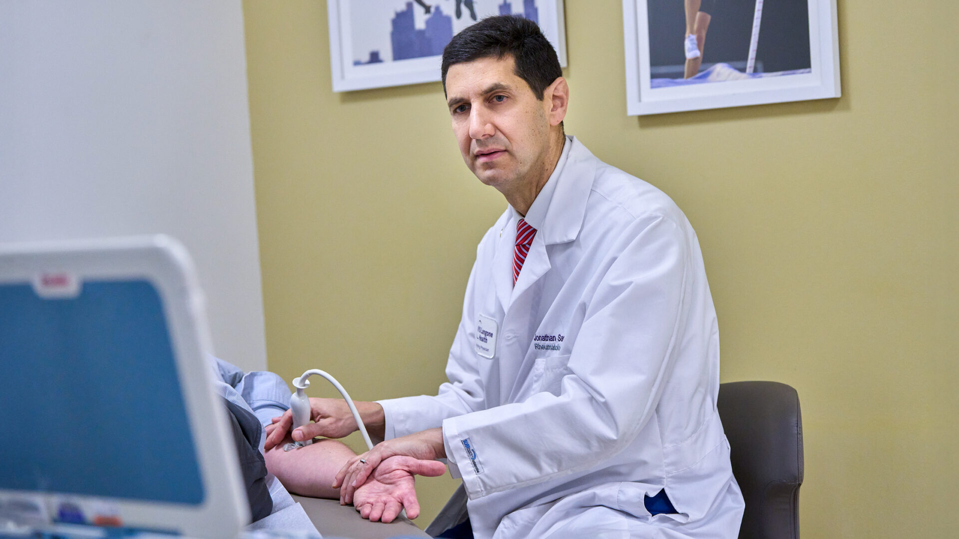
Moreover, the applications of MSK-US are remarkably diverse. In initial diagnosis, for example, US can identify gout and other crystal diseases, distinguish between arthritis types, and facilitate early detection of erosions, synovitis, and enthesitis. Subsequent findings can help guide treatment by demonstrating disease progression and remission. “We think of this technology as our version of the stethoscope,” Dr. Samuels says. “You can see how much fluid is in the knee during a clinical exam, and often identify other tears and accumulations, instead of scheduling an MRI and waiting for the results.”
MSK-US is also integral to image-guided aspiration and injection of nearly any joint from head to toe. Newer portable, handheld systems that connect wirelessly to mobile devices enable NYU Langone rheumatologists to take this approach beyond the outpatient exam room and onto the hospital wards for inpatient consultations.
“Being able to perform imaging and show it to the patient in a bedside consult is especially helpful when quick decisions must be made,” Dr. Samuels explains. “We can say, ‘here’s why we advise aggressive treatment,’ or ‘you’d be a good candidate for a particular clinical trial,’ and illustrate what’s happening in real time.”
Training New Generations of Experts
In addition to spearheading the use of MSK-US in rheumatology, NYU Langone is at the forefront of teaching clinicians to employ it. Since 2012, Dr. Samuels has directed NYU Langone’s annual two-day training course, now with beginner and intermediate tracks, which attracts about 50 physicians and allied health professionals from across the country to the NYU Langone campus each March.
“We’re proud to have helped spread this knowledge far beyond our own campus.”
Jonathan Samuels, MD
“Some participants have come back to teach the course, alongside seasoned specialists from NYU Langone and elsewhere,” Dr. Samuels notes. “Others go on to teach fellows at their own institutions. We’re proud to have helped spread this knowledge far beyond our own campus.”
That commitment to education also extends to national leadership in rheumatology US training. Dr. Samuels is vice president and rising president of the Ultrasound School of North American Rheumatologists (USSONAR), which now trains about 90 fellows and attendings each year through online modules and image uploads. The year-long USSONAR curriculum also includes a mandatory three-day, in-person course each January, which Dr. Samuels has directed since 2024.
Improving the Assessment of Fracture Risk
In osteoporosis, one of the most common comorbidities of rheumatic disease, dual-energy X-ray absorptiometry (DXA) has played a similarly transformative role, with newer imaging tools such as trabecular bone score (TBS) and high-resolution peripheral quantitative computerized tomography (HRpQCT) adding further refinements. NYU Langone rheumatologists were early adopters of the latter techniques, and are now advancing their use to improve the treatment of complex osteoporosis.
NYU Langone’s commitment to bone health dates back decades. The Osteoporosis and Bone Health Program, established in 1994 by renowned expert Stephen Honig, MD, has been recognized as an American Orthopedic Association “Own the Bone Star Performers” site for nearly 10 years.
Today, the program is led by Nicole Leung, MD, who trained under Dr. Honig as a rheumatology fellow at NYU Langone. “We hope to build upon the strong legacy of the Osteoporosis and Bone Health Program by continuing to optimize patient care and strengthening interdisciplinary collaboration,” says Dr. Leung. She completed additional training in clinical trial research methods and bioinformatics through NYU Langone’s Clinical and Translational Science Institute and is a certified clinical densitometrist.
As chief fellow for quality improvement, Dr. Leung led initiatives to optimize patient care and safety—a focus she has maintained since being appointed director of the osteoporosis program in 2024. One of her first initiatives was to develop an expedited post-fracture appointment protocol for patients at high risk of recurrent fractures. “We want to ensure those patients receive preventive interventions before they sustain another break,” she says. The clinic works closely with orthopedic surgeons, primary care physicians, and physical therapists to provide comprehensive and longitudinal care.
Since the 1990s, the gold standard technology for assessing fracture risk has been DXA, which uses X-rays at two distinct energy levels to determine bone mineral density (BMD). However, DXA imaging cannot detect changes in skeletal microarchitecture, which may predict bone fragility independently of areal BMD. Nor can it differentiate cortical from trabecular bone. Its reliability in fracture-risk assessment may be further impaired by only providing a two-dimensional measurement of a three-dimensional (3D) structure. For these reasons, Dr. Leung and colleagues at NYU Langone sometimes turn to TBS and HRpQCT.
Moving Beyond the Gold Standard
TBS gives additional information on the quality of the microarchitecture. Using software installed on a standard DXA computer, it generates an index of bone microarchitecture based on the texture of trabecular bone at the lumbar spine. While less detailed than HRpQCT, it has been shown to enhance prediction of fracture risk when used as a complement to DXA.
HRpQCT is a 3D imaging technique designed to evaluate bone microstructure as well as BMD, using a low-dose, high-resolution, quantitative CT scan. Studies show that this technique—the first to enable measurement of bone quality without a biopsy—can predict fracture risk more accurately than DXA.
“To find the right treatment, you need the right diagnosis, and using the right imaging method is essential.”
Nicole Leung, MD
“We use these newer techniques in complex patients who have artifacts that might lessen the accuracy or utility of bone-density measurements,” says Dr. Leung. “To find the right treatment, you need the right diagnosis, and using the right imaging method is essential.”