
Intertrochanteric femur fractures are among the most common of all fractures, especially in older individuals. These fractures are associated with substantial mortality and loss of mobility and generally require surgical treatment within 24 to 48 hours of admission, once the patient is medically optimized.1, 2, 3 Fracture nonunion is uncommon, with a reported incidence of 1 to 2 percent in all cases.4, 5 Reverse obliquity or transverse fractures are at higher risk for nonunion due to powerful deforming forces, the loss of lateral wall support, and exposure to shear forces that make reduction and fixation more difficult.6, 7, 8, 9, 10 In fact, the reported nonunion rate for reverse obliquity fracture patterns has been as high as 23 percent, more than 10 times higher than a standard obliquity fracture.6
Patients with a fracture nonunion often present with persistent hip or thigh pain, difficulty bearing weight, progressive functional decline, and loss of mobility; the nonhealing fracture also often results in varus deformity, implant failure, and the need for additional surgery.11, 12 Treatment with reconstructive surgery must address both the mechanical and biological causes of nonunion. In active patients with a viable femoral head, adequate proximal bone stock, and preserved articular cartilage, femoral head-preserving surgery is generally preferred. This approach typically involves removal of failed implants, debridement of the nonunion site, correction of the deformity, compression of the nonunion, and stable internal fixation. Autologous bone grafting may be beneficial in some cases.11, 12, 14, 15, 16
Conversion to arthroplasty is often performed for older, less active patients with severe osteoporosis, poor bone stock, irreparable damage to the femoral head or acetabulum, or advanced arthritic changes.11, 12, 13, 14, 15, 16, 17 While conversion arthroplasty may provide pain relief and facilitate mobilization, the procedure is much more technically demanding than primary joint arthroplasty and is associated with higher rates of infection, dislocation, and revision surgery, as well as worse overall functional outcomes.11, 17, 18, 19, 20
Here we present a case of an active older female who developed nonunion of an intertrochanteric femur fracture and was successfully treated with a valgus intertrochanteric osteotomy and blade plate fixation. The case highlights the complexity in decision making for treating intertrochanteric femur fracture nonunion and the ability to restore anatomy and function without conversion to complex hip arthroplasty.
Case Highlights:
- An 87-year-old female presented with persistent pain and loss of mobility after intramedullary nail fixation of a reverse obliquity intertrochanteric femur fracture.
- Imaging demonstrated medialization of the femoral shaft with shortening and varus alignment.
- Instead of conversion to complex hip arthroplasty, the patient was treated with an intertrochanteric osteotomy of the femur with blade plate fixation and compression through an articulated tensioning device.
- At 3.5 months postoperatively, X-rays revealed a healed fracture nonunion and the patient had no pain and could bear weight and ambulate without difficulty.
Case Presentation
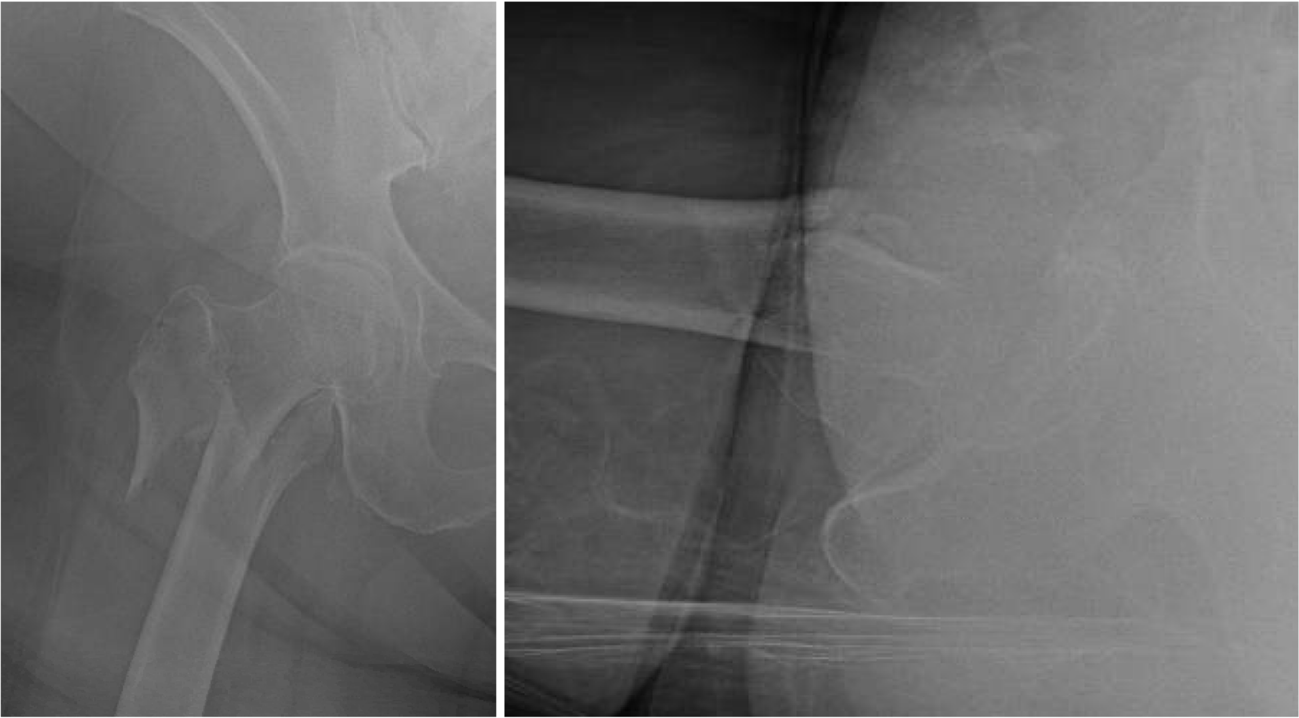
The patient is an 87-year-old female who sustained a low energy fall, could not get up, and was brought to the emergency department (ED). In the ED she complained of right hip pain. On exam, her hip was shortened and externally rotated, and any attempt at movement was painful. She remained neurologically intact, with no head strike or loss of consciousness. Plain X-rays, including an anteroposterior (AP) and cross-table lateral view of the right hip, demonstrated a displaced reverse oblique intertrochanteric hip fracture (Figure 1).
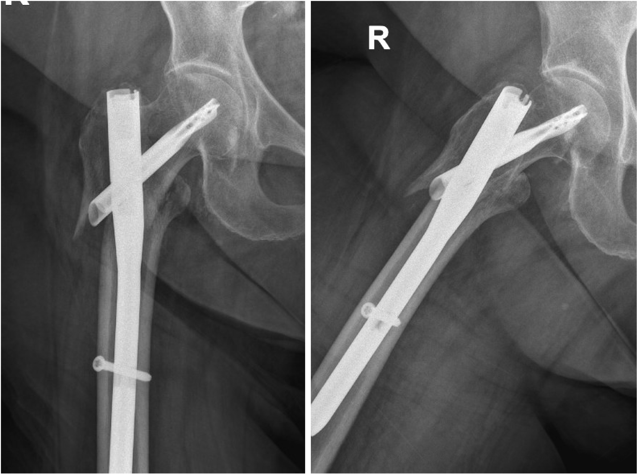
The patient was admitted to the hospital and underwent medical clearance. She had no significant medical history. She was a community ambulator, lived alone, and was active in her social life. On hospital day one, the patient underwent repair of the reverse obliquity fracture with a short cephalomedullary nail (Figure 2).
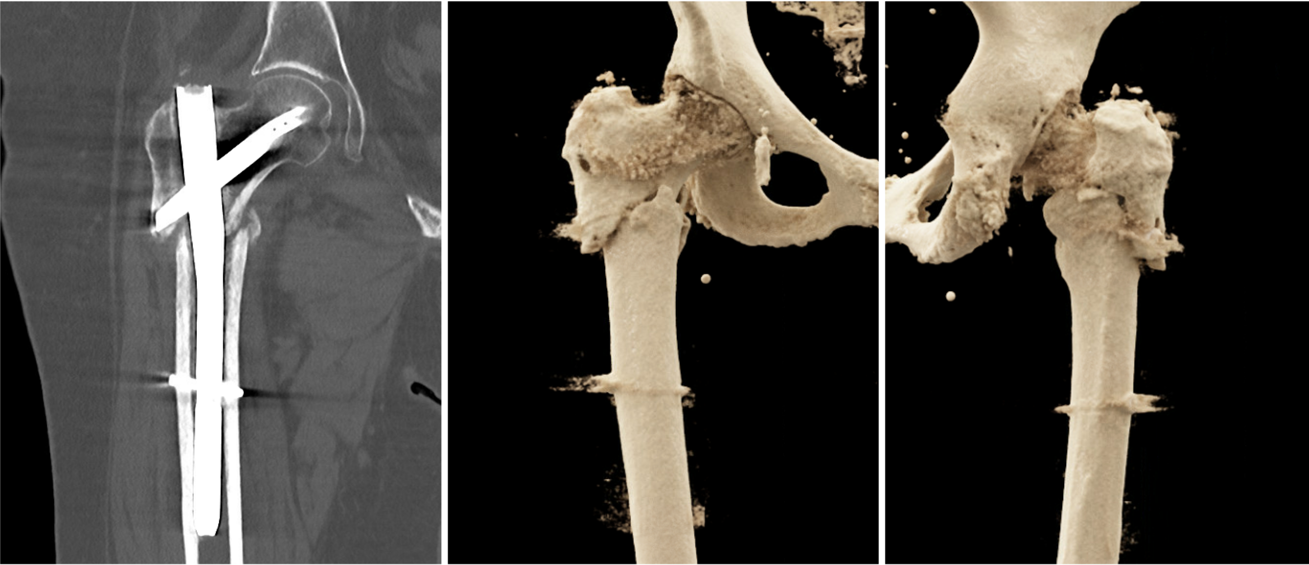
Her hospital course was unremarkable, and she was discharged to subacute rehabilitation and eventually to home. The patient, however, reported consistent pain in her thigh and right hip area and was unable to return to a normal gait. She had regularly scheduled follow-ups with her surgeon at 6 weeks and 3 months. At 4 months, with continued complaints of significant pain, a CT scan demonstrated incomplete healing of the fracture (Figure 3a). 3D-reconstructed CT scans showed some displacement of the shaft medially and shortening (Figure 3b,c).
At 6 months, the patient continued experiencing pain and was unable to walk independently. She was largely wheelchair bound and used a walker in the house. On physical exam, her initial surgical wounds were well healed, with no erythema or drainage. She had limited hip range of motion and 1-cm of shortening. A discussion was had with the patient regarding her surgical options, which included arthroplasty or hip preservation. Given the fact that her reverse oblique fracture would require a more complex hip replacement, a decision was made to proceed with hip joint salvage via osteotomy and nonunion repair. A preoperative infection lab screen was negative.
Operative Management
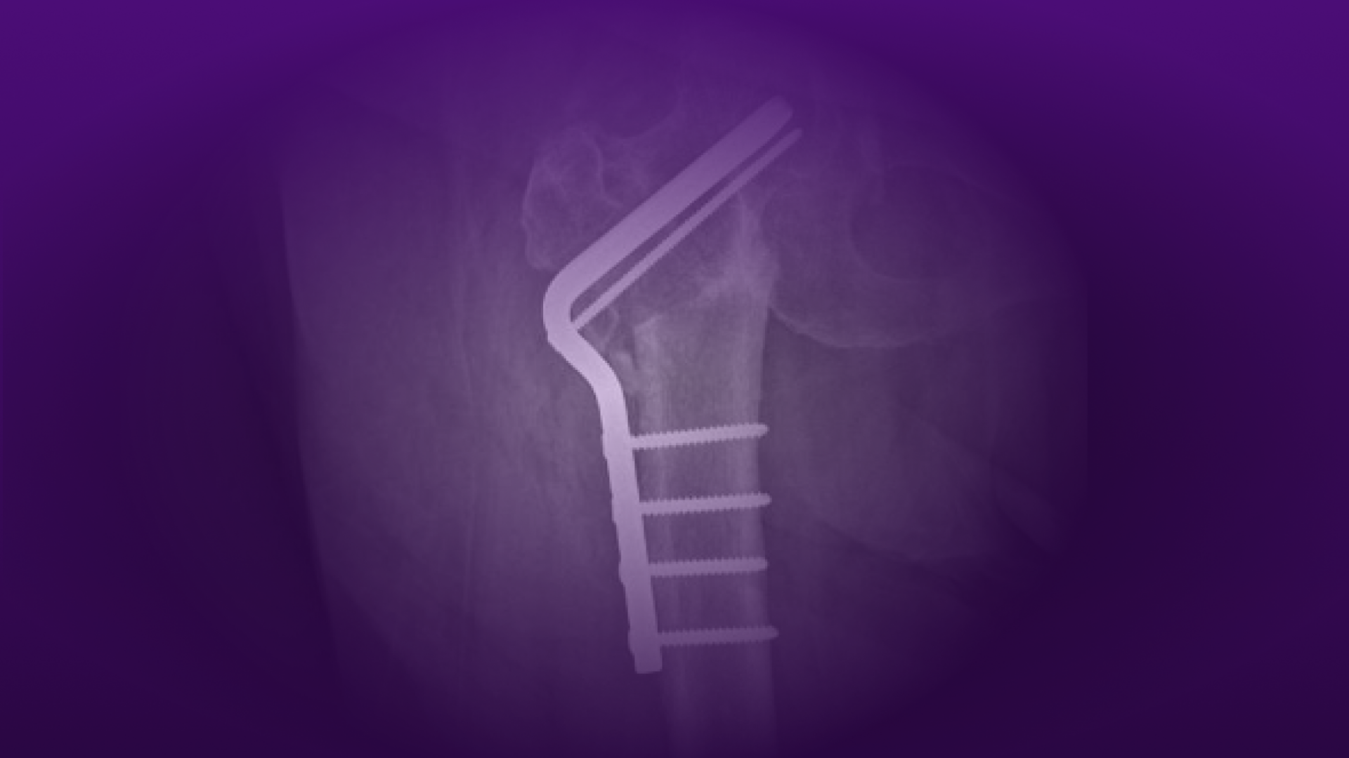
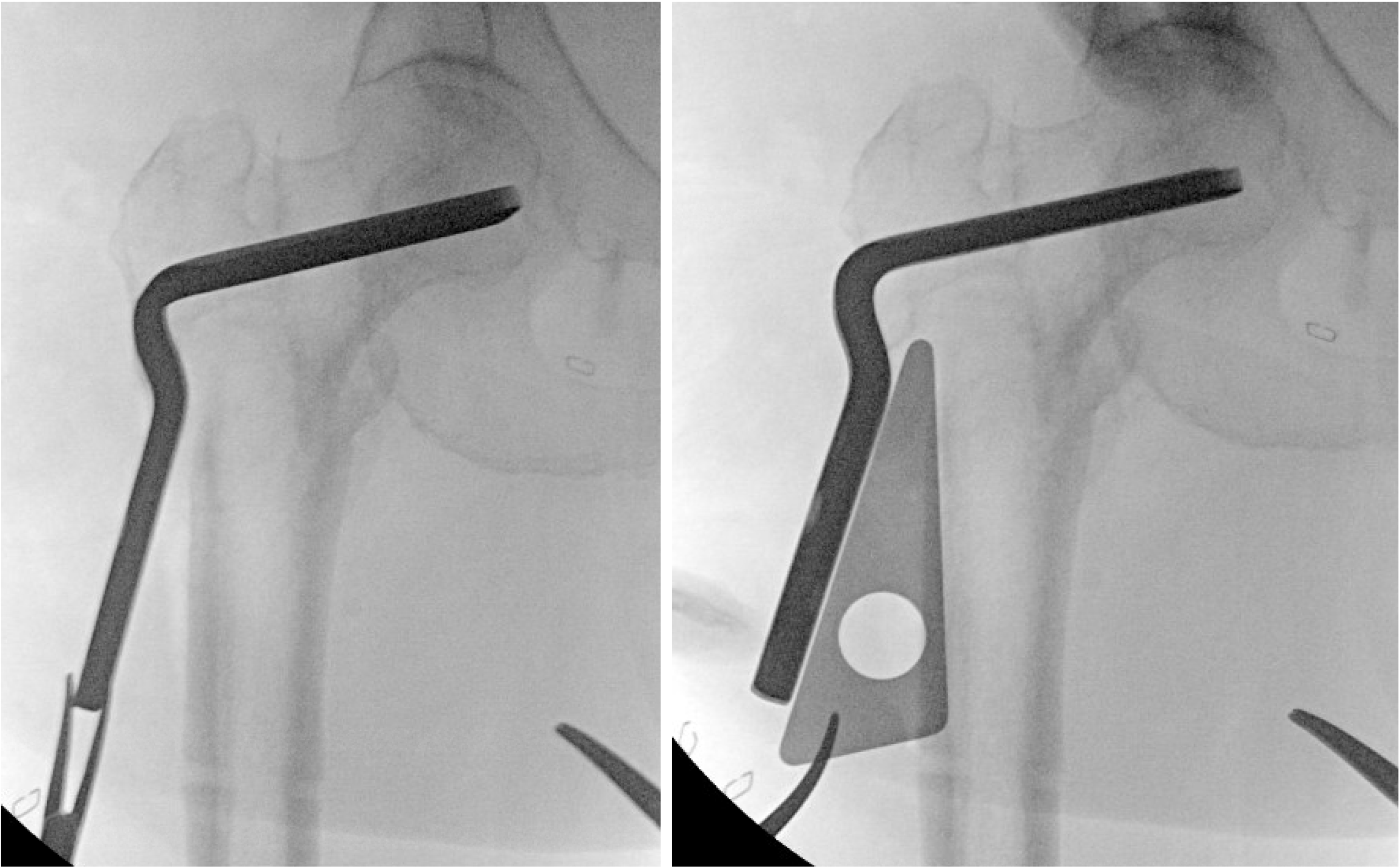
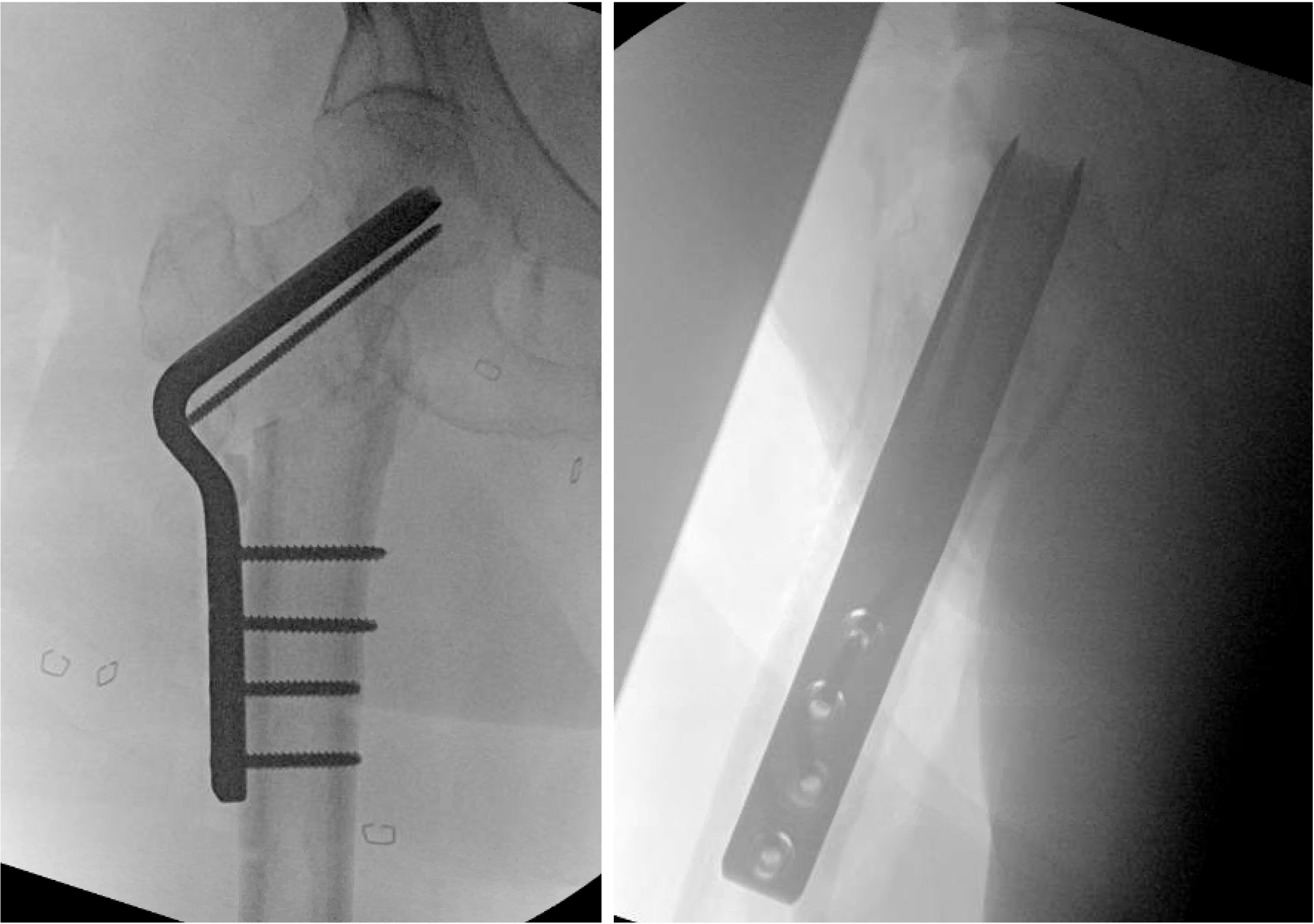
The patient was placed on a radiolucent flat table. A large lateral incision was made for a planned Pauwels’ osteotomy. Based on templating of the contralateral side, an intertrochanteric osteotomy was planned out with a 30-degree wedge and a 120-degree osteotomy plate to restore normal anatomy (Figure 4).
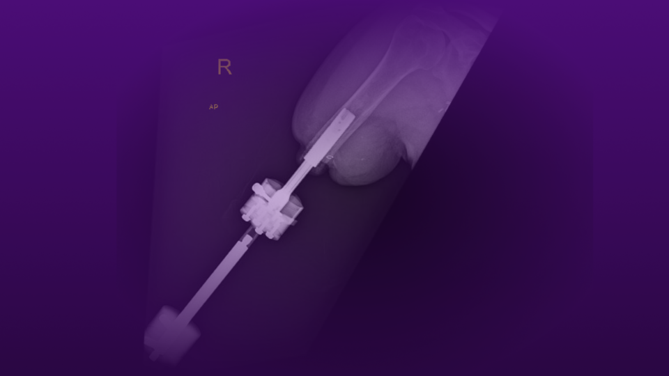
Surgery was performed under regional anesthesia with minimal blood loss. A bone wedge resection through the nonunion site left two fresh, flat bony surfaces for compression, which was achieved using the external articulated tensioning device and the ramped oval holes in the blade plate. Postoperative films are demonstrated in figure 5. The patient was discharged, again to subacute nursing, and achieved partial weight bearing.
Postoperative Course
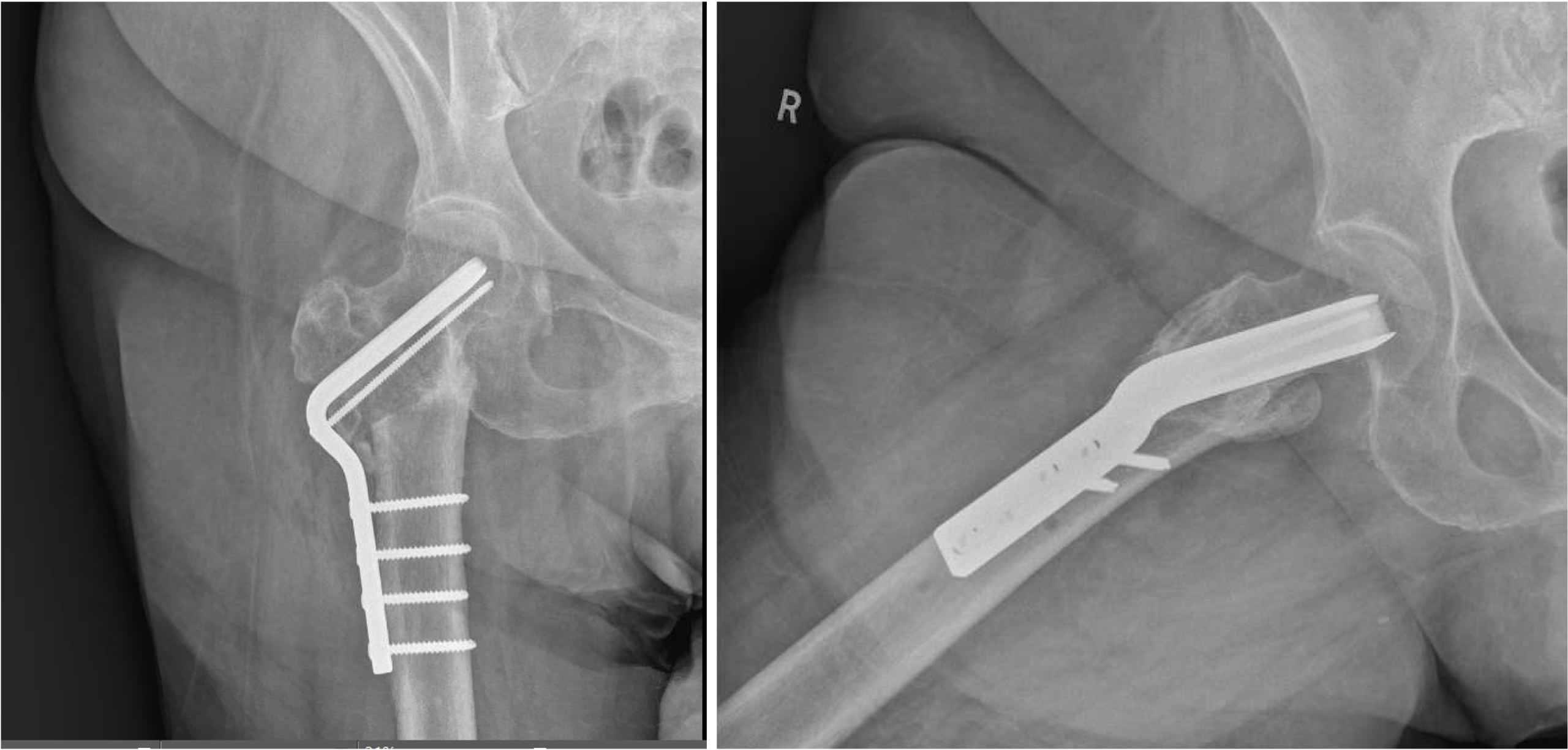
At 6 weeks post-surgery, X-rays demonstrated good alignment and early healing, and the patient advanced to full weight bearing, as tolerated. When she was seen again at 3.5 months, she had no pain and was able to fully bear weight (Video 1). X-rays, including an AP and lateral view of the right hip, demonstrated a healed subtrochanteric osteotomy with restoration of the normal neck shaft angle and limb length (Figure 6).
Discussion
Although rare, nonunion following surgery for an intertrochanteric femur fracture can be debilitating and lead to persistent pain and loss of mobility.11, 12 Radiographs demonstrate lack of callus formation with a visible fracture line. Progressive varus collapse and shortening of the fracture may be present along with implant loosening or implant breakage.10, 11, 12 Workup should include an assessment of the fracture alignment and implant stability as well as the amount of available bone stock for reconstruction and the viability of the femoral head. Infection must always be considered and ruled out. Modifiable risk factors, such as smoking, malnutrition, vitamin D deficiency, and other metabolic abnormalities also must be identified and addressed prior to any reconstructive surgery in order to increase the probability of a successful procedure. One recent study reported a 26.5 percent failure rate of operatively treated pertrochanteric fracture nonunions, with the failure rate reaching 50 percent in patients who are actively smoking or with medial bone loss.13
Treatment options for hip fracture nonunion consist of femoral head-preserving procedures and conversion to hip arthroplasty. While conversion to total hip arthroplasty remains a viable option for a significant number of failed hip fracture surgeries, this case represents an opportunity for hip preservation and demonstrates how an intertrochanteric osteotomy, using a blade plate and screw construct, can restore a patient’s normal gait and hip function, regardless of age. The above video demonstrates the patient’s gait and satisfaction 3 months after the procedure.
Conversion hip arthroplasty in the setting of intertrochanteric hip fracture nonunion is associated with higher rates of prosthetic joint infection (PJI), dislocation, and limb length discrepancy than primary hip arthroplasty.11, 17, 18, 19, 20 Compared to femoral neck fracture nonunion converted to hip arthroplasty, extra-capsular intertrochanteric fractures are often associated with lateral wall and/or calcar deficiency, subtrochanteric extension, proximal femoral deformity, greater trochanteric displacement, abductor dysfunction, and stress risers from previous internal fixation. This can result in a significantly longer operative time, increased blood loss, and increased risk of periprosthetic fracture.21, 22
Outcomes after conversion arthroplasty for extra-capsular hip fracture nonunion are generally worse, with more chronic pain, than for femoral neck fracture nonunion and are more comparable to revision hip arthroplasty than to primary total hip arthroplasty.23, 24 In comparison to standard-obliquity intertrochanteric fractures, reverse-obliquity fractures have greater deforming forces, greater proximal femoral deformity, and less viable proximal bone stock, all of which make arthroplasty particularly challenging.7, 8, 25
Prior to sustaining her hip fracture, the patient in this case was active with no complaints, and an intertrochanteric osteotomy with blade plate fixation restored her pre-injury function without a conversion to arthroplasty. In this scenario, conversion to arthroplasty likely would have required long diaphyseal fixation with cable or plate-augmented fixation of the trochanters or conversion to a proximal femoral replacement—strategies less likely to have achieved the positive result seen here.
References
- Forte ML, et al. J Bone Joint Surg Am. 2010;92(4):799–806. DOI.
- Dyer SM, et al. BMC Geriatr. 2016;16:158. DOI.
- Brox WT, et al. J Bone Joint Surg Am. 2015;97(14):1196–1199. DOI.
- Giordano V, et al. BMC Musculoskelet Disord. 2022;23:225. DOI.
- Walter N, et al. Bone Jt Open. 2023;4(10):801-807. DOI.
- Berk T, et al. BMC Geriatr. 2022;22:990. DOI.
- Haidukewych GJ, Israel TA, Berry DJ. J Bone Joint Surg Am. 2001;83(5):643-650. DOI.
- Sadowski C, et al. J Bone Joint Surg Am. 2002;84(3):372-381. Link.
- Sekimura T, Son SJ, Lee C. J Orthop Trauma. 2023;37(10S):S19-S25. DOI.
- Palm H, et al. J Bone Joint Surg Am. 2007;89(3):470-475. DOI.
- Angelini M, et al. J Orthop Trauma. 2009;23(6):471-478. DOI.
- Githens M. J Orthop Trauma. 2023;37(10S):S11-S18. DOI.
- Novak AA, et al. J Orthop Trauma. Published online May 13, 2025. DOI.
- Dhammi IK, et al. Indian J Orthop. 2011;45(6):514-519. DOI.
- Lim EJ, et al. Injury. 2021;52(10):2787-2794. DOI.
- Min BW, et al. Injury. 2019;50(7):1339-1346. DOI.
- Liu P, et al. BMC Musculoskelet Disord. 2020;21:573. DOI.
- Smith A, et al. Bone Joint J. 2019;101-B(6 Suppl B):91-96. DOI.
- Douglas SJ, et al. 2021;36(7 Suppl):S155-S159. DOI.
- Biniam B, et al. Arthroplast Today. 2025;33:101715. DOI.
- Mortazavi SMJ, et al. J Arthroplasty. 2012;27(1):31–36. DOI.
- Archibeck MJ, et al. J Arthroplasty. 2013;28(1):168–171. DOI.
- Hung CC, et al. J Orthop Surg Res. 2023;18(1):45. DOI.
- Tahir M, et al. J Clin Orthop Trauma. 2026;81:103542. DOI.
- Haidukewych GJ, Berry DJ. Clin Orthop Relat Res. 2003;412:184–188. DOI.