

Traumatic talus dislocations are among the most challenging injuries encountered in orthopedic trauma surgery because of the talus’s unique anatomy, limited vascular supply, and critical role in ankle and hindfoot biomechanics. Complete talar extrusion or non-reconstructable talar destruction is exceptionally rare and is often associated with high-energy trauma, substantial soft tissue compromise, and a high risk of complications including avascular necrosis, infection, post-traumatic arthritis, and long-term functional disability. Preservation of the native talus is generally preferred when feasible; however, in cases in which no viable talar bone remains intact, reconstructive options become limited and technically demanding.1,2,3
Historically, treatment strategies for severe talar injuries have included talectomy with tibiocalcaneal arthrodesis, bulk allograft reconstruction, or staged salvage procedures, each of which carries substantial morbidity and often results in impaired hindfoot motion and altered gait mechanics. In recent years, total talus implantation has emerged as a promising limb-salvage technique for patients with irreparable talar loss or collapse. Advances in three-dimensional (3D) imaging and custom implant manufacturing have enabled the development of patient-specific metallic total talus prostheses designed to restore native anatomy, preserve limb length, and maintain ankle and subtalar joint congruity. Early reports demonstrate encouraging outcomes with improved pain control, functional recovery, and maintenance of mobility compared with traditional fusion procedures.1,2,4
Despite growing interest in total talus replacement, the literature remains limited largely to small case series and reports, particularly in the setting of acute traumatic talus extrusion or complete talar loss. Management of these injuries remains difficult because of the rarity of the condition, lack of standardized treatment algorithms, challenges in soft tissue management, and uncertainty regarding long-term implant survivorship and complication rates. Furthermore, surgeons must balance the goals of restoring function and preserving motion against the risks of infection, implant failure, instability, and adjacent joint degeneration.3,5,6
Here we present a case of a traumatic talus fracture dislocation with associated Lisfranc fracture successfully treated with staged surgical procedures and ultimate total talus replacement.
Case Highlights:
- A 22-year-old male presented with a traumatic extruded talus fracture dislocation and associated Lisfranc fracture, with a 5-cm lateral hindfoot laceration and severe talar bone loss (<40 percent of the talus remaining, including complete loss of the head and neck).
- Initial management included irrigation and debridement with application of a spanning external fixator.
- Approximately 2 months later, he underwent definitive reconstruction with total talus implantation and arthrodesis of the Lisfranc complex and midfoot.
- At 1-year follow-up, the patient ambulates comfortably and maintains 10 degrees of both plantarflexion and dorsiflexion at the ankle joint.
Case Presentation
The patient is a 22-year-old male with no significant past medical history, who presented to the emergency department (ED) with an extruded talus fracture dislocation and associated Lisfranc fracture. The patient was found unresponsive with isolated severe left foot pain.
On examination, he had a 5-cm laceration over the lateral aspect of the hindfoot without pulsatile bleeding. He was neurovascularly intact in the distal foot with strong pulses. He received antibiotics and tetanus prophylaxis on presentation to the ED and the limb was placed in a splint.
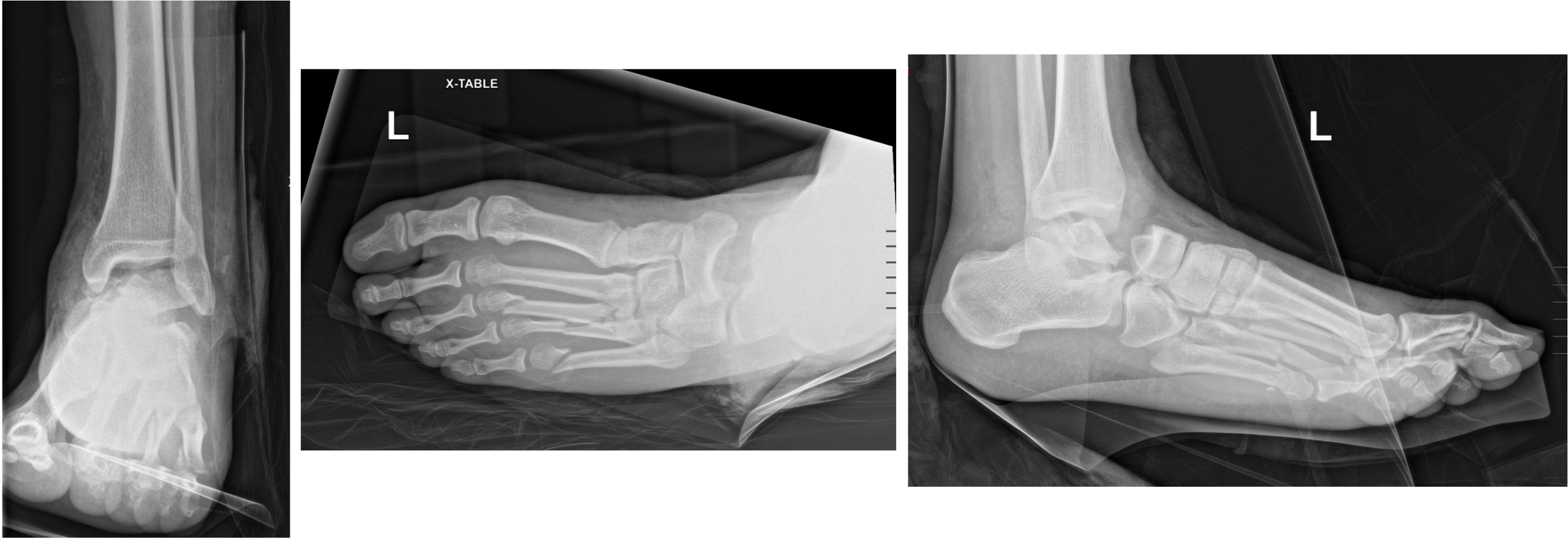
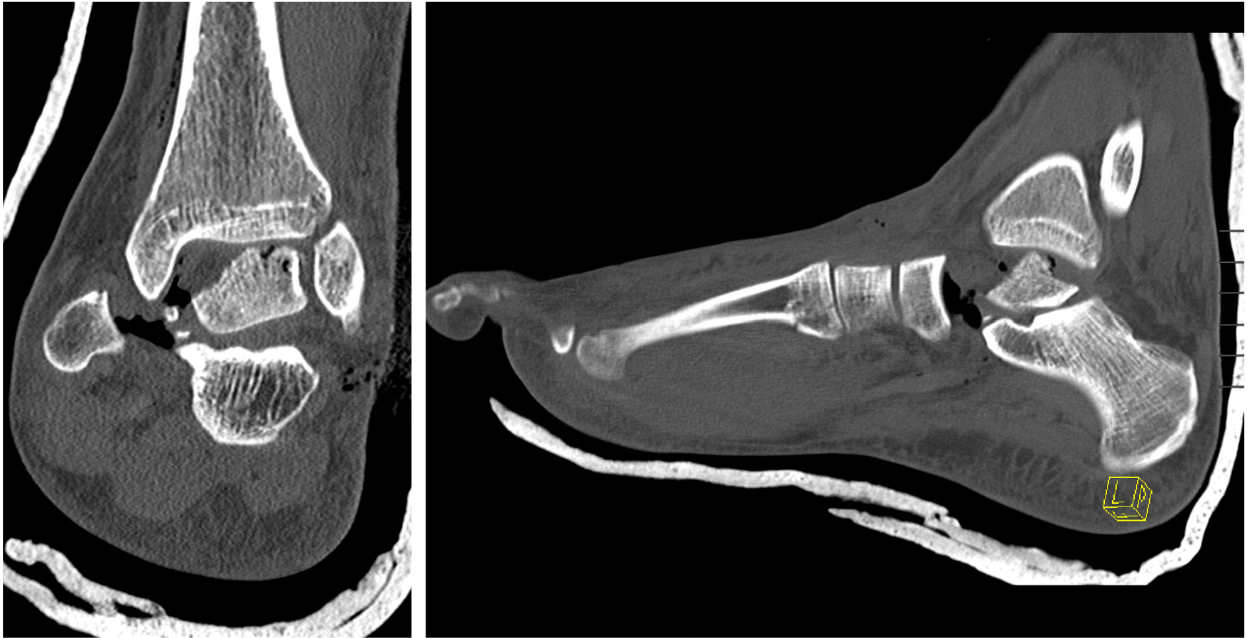
Radiographs and CT demonstrated fracture dislocation of the talus with extensive bone loss. Less than 40 percent of the talar body remained in situ and the talar neck and head were completely absent. There was also an associated Lisfranc fracture-dislocation involving fractures of the third through fifth metatarsals (MTs) (Fig 1 and 2).
The devastating nature of the injury was discussed with the patient, and he was indicated for staged surgical management.
Operative Management
Initial Operative Intervention. The patient was brought to the operating room (OR) within 6 hours of presentation to the hospital. He was placed supine on an OR table and the leg was prepped and draped.
A thorough exploration of the wound and residual talus was performed. The residual talus comprised less than 40 percent of the talar body, lacked soft tissue attachments, and had significantly injured cartilage. The decision was made to discard it due to risk of infection and necrosis. The wound was then copiously irrigated and closed and a spanning external fixator was applied, utilizing a standard delta frame, extending into the foot to maintain length (Fig 3). Finally, the third through fifth MTs were pinned utilizing Kirschner wires.
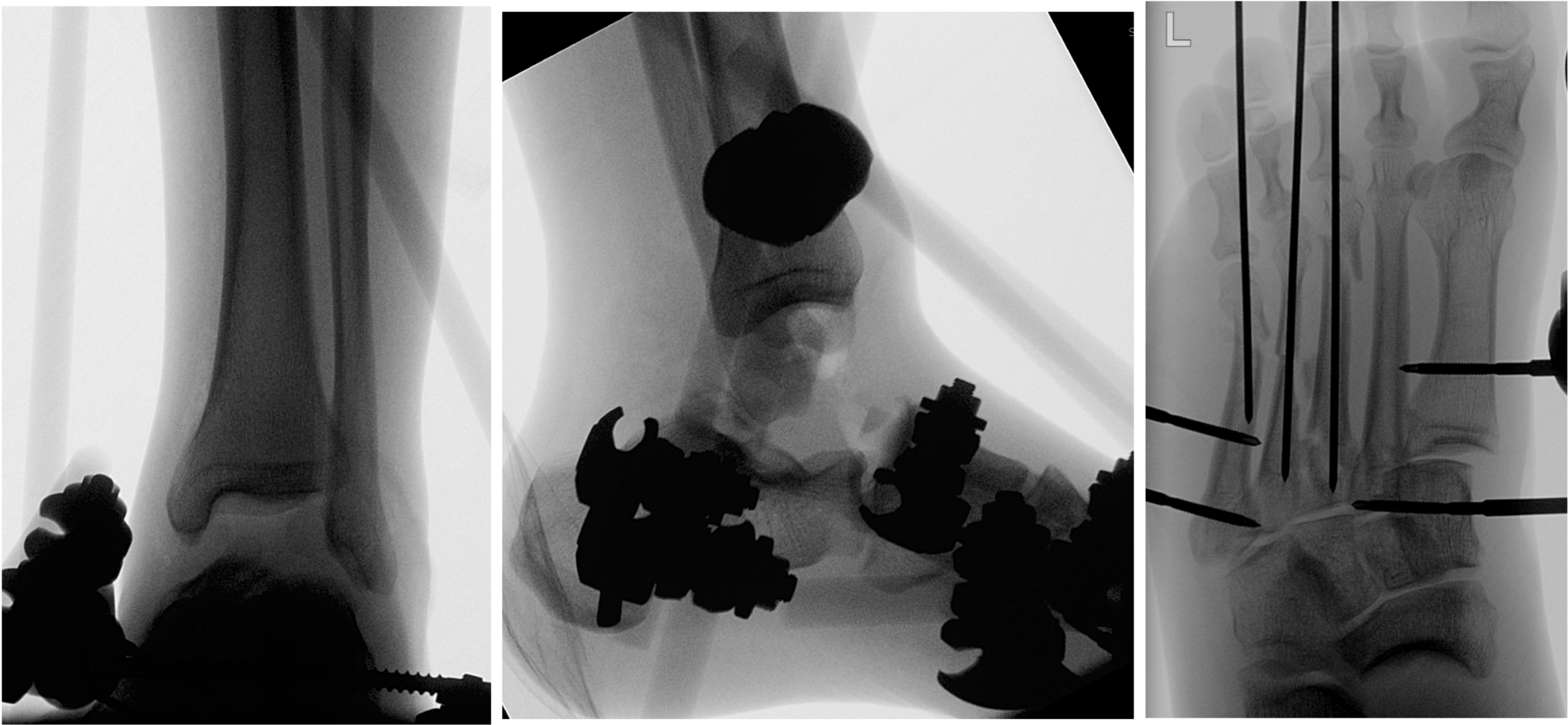
Initial Postoperative Course. The patient was admitted for 48 hours of antibiotics for open fracture. He was made non-weight-bearing and CT scans of the bilateral ankles were obtained to plan for a total talus replacement.
Second Operative Intervention. Approximately 2 months after his index procedure, the patient was returned to the OR for definitive management of his injuries. He was placed supine on the OR table and the external fixator was removed as well as the metatarsal pins.
A midline longitudinal incision was made directly over the anterior aspect of the ankle joint extending slightly medial towards the midline of the navicular. Once entering the talus joint space, there was a significant amount of scar tissue which was then sharply debrided. The underlying cartilage of the tibial plafond as well as subtalar facets of the calcaneus were maintained and some small remaining talar bone fragments were removed along the posterior medial aspect of the joint space.
The medium size talar trial was then inserted into the space and felt to be the appropriate size using anteroposterior (AP) and lateral fluoroscopy to confirm positioning. At this point, the custom-made medium size total talar implant was carefully inserted into the joint space. Once implanted, that ankle was brought through range of motion and shown to be a good fit through plantarflexion and dorsiflexion. Eversion and inversion were appropriate through the subtalar joint. Final AP and lateral radiographs were obtained to confirm the position and placement of the total talar implant.
Once the talus replacement had been completed, attention was turned to the Lisfranc fracture-dislocation. The incision for the talus was extended to reach the Lisfranc joint and a secondary incision was made over the third tarsometatarsal (TMT) joint. The cartilage was removed with a combination of burr and curettes. A reduction of the Lisfranc joint and third TMT was then performed and held in place with Kirschner wires. The Lisfranc arthrodesis and third TMT fracture open reduction and internal fixation (ORIF) was then performed utilizing a combination of lag screws and plates.
Final fluoroscopic imaging demonstrated appropriate restoration of the arch of the foot and placement of all hardware (Fig 4). The patient was placed in a short leg splint postoperatively.
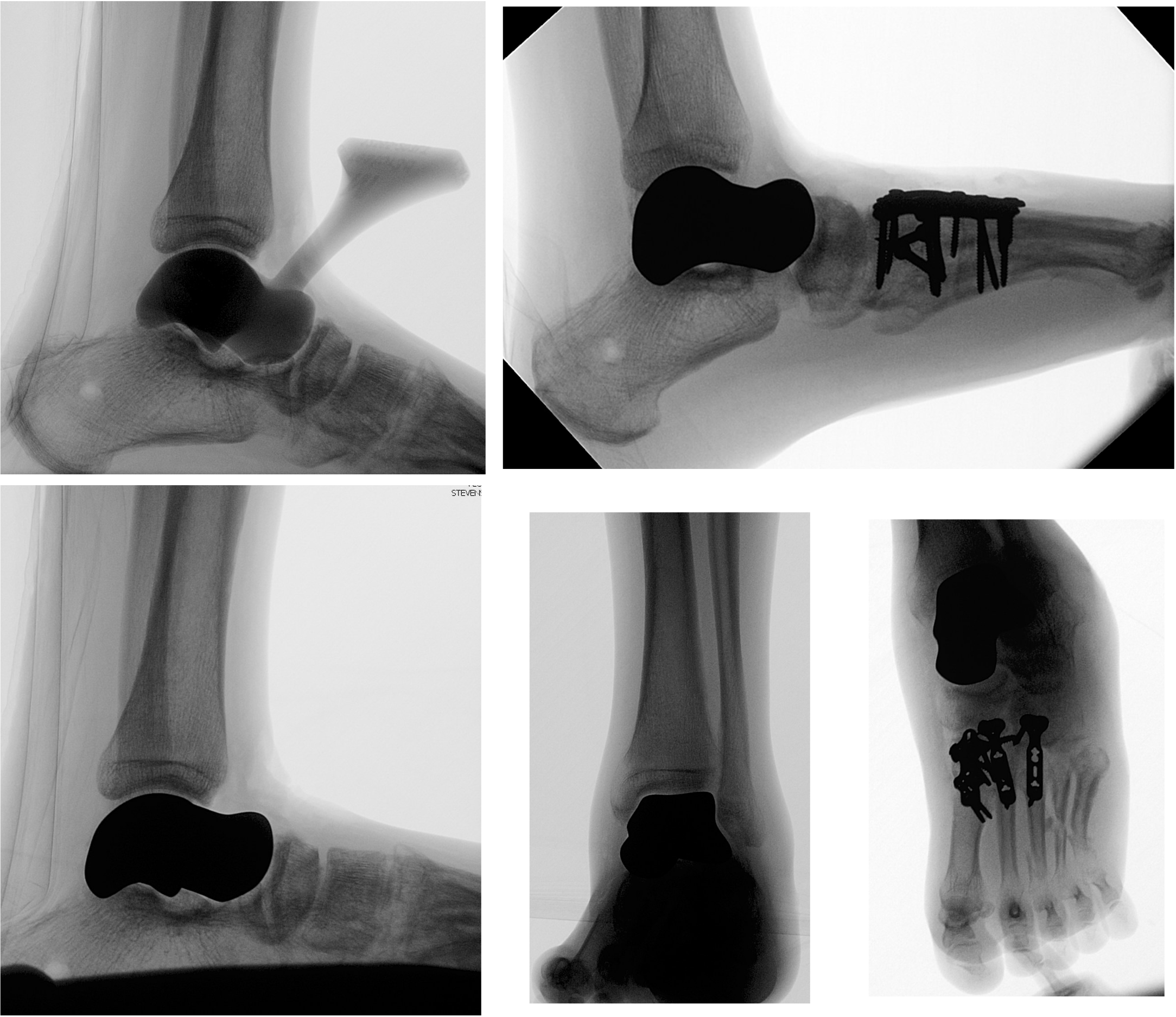
Postoperative Course
The patient was made non-weight-bearing for 3 months. He was allowed to initiate motion at 4 weeks following a period of soft tissue rest.
He was seen most recently at 1 year postoperatively. At that time, he was overall very happy with his foot. He was able to walk comfortably with a cane in the community and without a cane around the house. He had start-up pain at the first 5 to 10 minutes after sitting for long periods. He had returned to work. On exam, his incisions were well healed. His ankle range of motion was dorsiflexion 10 degrees and plantarflexion 10 degrees. He walked with a slightly stiff gait and shortened stance phase. Radiographs demonstrated overall acceptable alignment without hardware complications or tibial plafond arthritis (Fig 5).
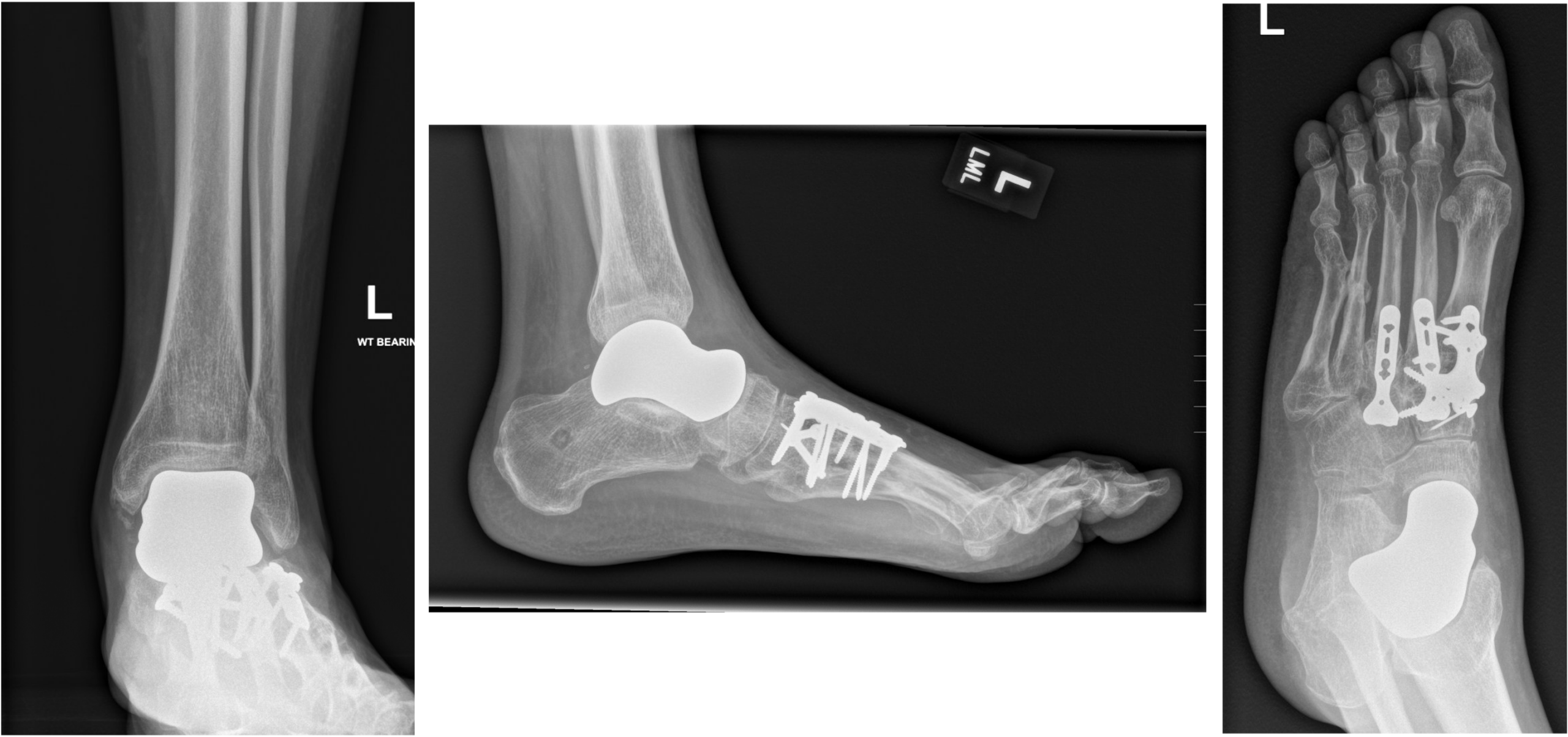
Discussion
Extruded talus fracture-dislocations are an extremely rare and difficult orthopedic injury, constituting only about 2 percent of talus fractures, which in themselves are rare.1,7 Treatment guidelines remain controversial as to whether the extruded talus should be reimplanted, replaced or if the hindfoot should be fused.
Several small case series demonstrate favorable outcomes with urgent (less than 3 to 6 hours) reimplantation of the talus, although complications are not uncommon, including infection, avascular necrosis, and collapse.2,4,7,8,9 Some authors advocate for reimplantation even in late cases, as it can be useful for bone stalk if the need arises for fusion.
In our case, where the extruded talus was not available, the only viable options were talus replacement or arthrodesis (tibiocalcaneal or allograft bone block).
Historically, extruded talus injuries were treated with the Blair fusion technique, a form of tibiocalcaneal arthrodesis. However, this technique shortened the affected limb leading to significant complications and limited function for the patient.2,5,10 Our patient also had an adjacent Lisfranc injury; a hindfoot arthrodesis with an associated midfoot arthrodesis would lead to very poor function of the foot and difficulty with ambulating.
Total talus replacement is a relatively new surgical procedure which relies on patient-specific custom implant generation utilizing 3D printing technology. It is indicated as a salvage procedure in talar extrusion, high-grade avascular necrosis of the talus without significant tibiotalar arthritis, and for large talar lesions including tumors. Several small case reports and series have demonstrated excellent outcomes, with some patients even returning to sport.5,11
As the procedure remains new, there is limited long-term data. However, several authors have reported on complications, including subsequent tibial plafond arthritis and need for conversion to ankle replacement or arthrodesis.5 In our young patient, with a multiply injured foot and need for midfoot arthrodesis, the talar replacement was an excellent option for preserving motion.
In summary, talar extrusion is a rare and challenging injury. If the talus is available, it should likely be reimplanted unless there are extenuating circumstances. If the talus is lost, total talus replacement is a viable option and has been found, in limited series, to have good outcomes.
References
- Van Opstal N, Vandeputte G. Acta Orthop Belg. 2009;75(5):699-704. DOI. Link.
- Weston JT, et al. Orthop Surg. 2015;7:97–101. DOI.
- Johnson LG, et al. Foot Ankle Surg. 2022;28(8):1194-1201. DOI.
- Kwak JM, Heo SK, Jung GH. J Med Case Rep. 2017;11(1):348. DOI.
- Leonetti D, et al. Medicina (Kaunas). 2023;59(8):1498. DOI.
- Anastasio AT, et al. Foot Ankle Spec. 2025;18(4):415-428. DOI.
- Gupta TP, et al. J Orthop Case Rep. 2024;14(1):5-10. DOI.
- Memisoglu K, Hürmeydan A. J Am Podiatr Med Assoc. 2009;99(5):431-434. DOI.
- AlMaeen BN, et al. Cureus. 2020;12(5):e7947. DOI.
- Vaienti L, et al. J Orthop Traumatol. 2011;12(1):61-64. DOI.
- Katsui R, et al. Foot Ankle Int. 2020;41(1):79-83. DOI.