
Referral Notes:
- NYU Langone’s Microtia Services for Children Program provides centralized services for hearing loss and external ear anomalies.
- The multidisciplinary team combines pediatric otolaryngology, facial plastic and reconstructive surgery, audiology, and otology under one roof.
- The program accepts patients from birth and manages a high volume of microtia referrals.
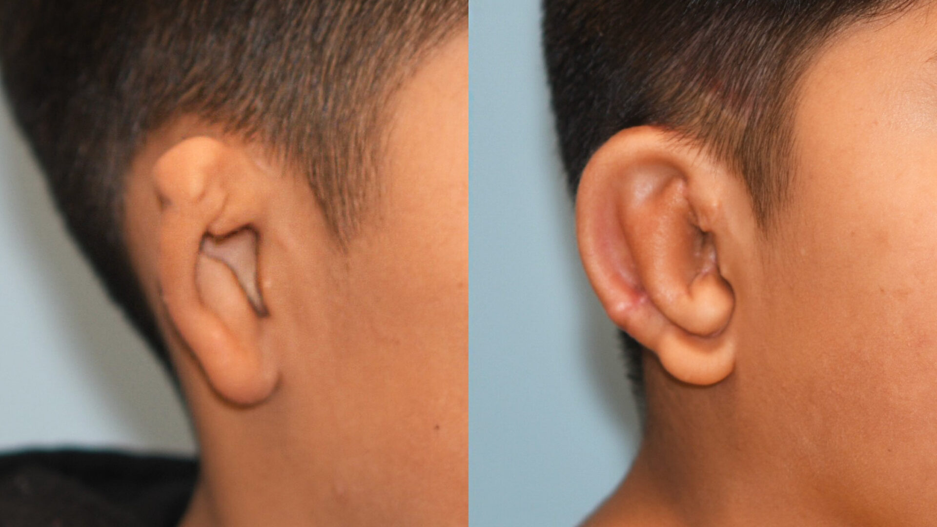
A boy younger than 10 years of age with microtia of the right ear was referred by an outside institution to the Microtia Services for Children Program at Hassenfeld Children’s Hospital at NYU Langone for specialized care. He used a bone-anchored hearing aid, and the external structure of his right ear had not undergone surgical reconstruction.
The dedicated microtia program—one of a limited number of multidisciplinary pediatric microtia programs in the US— delivers comprehensive care for children who require ear reconstruction and hearing management. The program is co-led by otolaryngologist and facial plastic surgeon Danielle Eytan, MD, and pediatric otolaryngologist Zahrah Taufique, MD, MBA. Neurotologist David R. Friedmann, MD, and audiologist Erin Byrd, AUD, round out the multidisciplinary team.
“We wanted to provide a better care experience for these patients and their families by creating a home for them, where they could get their care all under one roof on the same day.”
Danielle Eytan, MD
“Patients with microtia are extremely complex and require extensive preoperative and postoperative care with multiple appointments,” Dr. Eytan says. “We wanted to provide a better care experience for these patients and their families by creating a home for them, where they could get their care all under one roof on the same day.”
Although microtia is rare, affecting about 1 in every 3,800 babies in the US, the clinic sees approximately 10 patients and performs one to two surgical procedures each month.
Two-Stage Reconstruction
Reconstructive surgery for the external ear is typically performed around age 7, when patients have enough rib cartilage to create an ear framework to match the opposite ear and allow for optimal symmetry. The patient was still eligible for surgery even though he was a few years older. Until patients are old enough to be eligible for surgery, they are monitored closely and receive counseling, audiology services, and support for appropriate language development.
“Since the referred patient was already old enough for surgery, we immediately began evaluating him for rib cartilage graft reconstruction,” Dr. Eytan says.
A specialized CT scan revealed a narrowed ear canal that impaired hearing while confirming the presence of some inner ear structures. An audiology evaluation by Dr. Byrd indicated moderately severe conductive hearing loss, supporting a diagnosis of Grade 3 microtia.
The rib cartilage graft reconstruction was performed in two stages, with both surgeons operating in tandem. “Dr. Taufique and I do all of the surgeries together,” Dr. Eytan says. “This two-surgeon approach brings the expertise of pediatric ENT and facial plastic surgery to every case.”
“Our goal is to make the ear look as normal as possible so other kids don’t notice and the patient never has to think about their ear again.”
Zahrah Taufique, MD, MBA
During the first stage, Drs. Eytan and Taufique harvested cartilage from ribs six through nine, using scalpels to meticulously hand-carved the cartilage into the framework of the new right ear. The sculpted cartilage was then implanted beneath the skin on the affected side, creating an ear that mirrored the shape of the patient’s unaffected ear while lying against the skull.
“Although synthetic implants are an option, for most children we use rib cartilage,” Dr. Taufique says. “When we use a patient’s own tissue, the reconstructed ear is truly their own. The body accepts it naturally, it grows and heals as living tissue, and it’s more resilient to the bumps and scrapes of childhood.”
A few months later, after the overlying skin conformed to the structure and vascularization was established, the surgical team performed the second-stage surgery. During the procedure, the reconstructed ear was elevated to match the natural projection of the unaffected ear and securely positioned with additional cartilage placed beneath the skin.
“We want a harmonious ear that matches the face and the other side,” Dr. Taufique says. A scalp skin graft, harvested from a hair-bearing area to conceal the donor site, was used to create the postauricular sulcus.
Functional and Psychosocial Outcomes
“Research shows that patients with microtia can experience psychological challenges because of the deformity. They get teased, and they feel different,” Dr. Taufique says. “Our goal is to make the ear look as normal as possible so other kids don’t notice and the patient never has to think about their ear again.”
The second stage of surgery is also an opportunity to place a surgically implanted bone-anchored hearing aid. In this case, the patient was a candidate for an atresiaplasty due to their favorable CT scan (high Jahrsdoerfer score). Dr. Friedmann will perform the surgery in the near future, with the goal of allowing the patient’s hearing to be conducted through the native structures.
Meanwhile, with his new natural-looking right ear, “the patient and his family are very happy with the results,” Dr. Taufique says.